
Daily Clinical Briefing · Educational reading for healthcare professionals
An asymptomatic dilated ascending aorta needs whole-aorta imaging; sudden chest or back pain adds haemodynamic control and surgical discussion.
A dilated ascending aorta needs whole-aorta imaging before surveillance decisions.
Sudden severe chest or back pain with high blood pressure should keep acute aortic dissection in view until anatomy and haemodynamics are addressed. Aortopathy should come first because it links clinic surveillance with emergency dissection care: confirm echo dilatation with CT aortography or cardiac magnetic resonance imaging, document diameter, valve morphology, enlargement rate and high-risk features, then involve cardiothoracic surgery immediately when type A dissection is suspected.
ECMO in a cancer patient adds a critical-care escalation decision: malignancy is not an automatic exclusion, but the bridge, reversibility, treatment options and review point must be explicit. Gastroenteritis brings the ward and community check: hydration, isolation, stool testing triggers and antibiotic restraint. Prosthetic heart valves add a concise examination frame for breathlessness, anticoagulation and endocarditis screening.

Incidental ascending aortic dilatation or sudden chest/back pain needs more than echo alone. Confirm size with CT aortography or cardiac magnetic resonance imaging, map the whole aorta, and in suspected type A dissection control heart rate and systolic blood pressure while involving cardiothoracic surgery.

Vomiting and diarrhoea are common, but hydration risk, isolation and stool testing still need structure. Exposure timing can point to norovirus, Campylobacter, Bacillus cereus or giardiasis, while possible E. coli O157 makes antibiotic restraint clinically important.
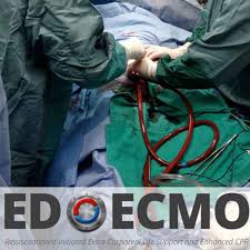
Cancer should not end an ECMO discussion by itself. Clarify the reversible problem, tumour type, treatment line, targetable mutations, immunotherapy toxicity and likely time to response, then define the bridge and review point with oncology and critical care together.

A metallic valve sound with a sternotomy scar should trigger early localisation and complication screening. Breathlessness after valve replacement needs valve function, ventricular function, rhythm, inflammation and anticoagulation reviewed rather than a purely descriptive examination summary.
When echocardiography reports ascending aortic dilatation, do not leave the next review vague. Arrange CT aortography or cardiac magnetic resonance imaging, record valve morphology, diameter, enlargement rate and blood pressure. The aortopathy topic links surveillance thresholds with dissection treatment.
What should follow an echocardiographic finding of ascending aortic dilatation?
Arrange CT aortography or cardiac magnetic resonance imaging to define the whole aorta. Document valve morphology, diameter, blood pressure, enlargement rate and high-risk features before deciding surveillance or surgical referral.
In suspected acute type A aortic dissection, what action should run in parallel with cardiothoracic discussion?
Start haemodynamic control with beta blockade and analgesia where appropriate. The stated targets are systolic blood pressure below 120 mmHg and heart rate below 60 beats per minute.
Before excluding ECMO because of cancer, what should be clarified?
Clarify the immediate reversible problem, treatment options, likelihood and time to response, and expected quality of life if the acute insult resolves. Agree the bridge, review point and criteria for continuing, escalating or withdrawing support.
Daily Clinical Briefings are prepared using ChatGPT Pro from the show notes and educational output for that day’s episodes. Iain Beardsell then checks the briefing for accuracy.
This describes the current briefing process. Item-specific review dates are shown only where documented. The original podcasts remain the sources; MedPod’s briefing is additional educational material.
Read about MedPod’s editorial approach
Report an error or source concern — please include the article title and link, without patient-identifying information.




.svg)